Pulmonary Carcinoma
Copyright Veterinaryradiology.net
Publication Date: 2014-01-02
History
12 year old male neutered Domestic Shorthair Cat with 2 week history of increased respiratory effort and several days of inappetence.
3 images
Thorax
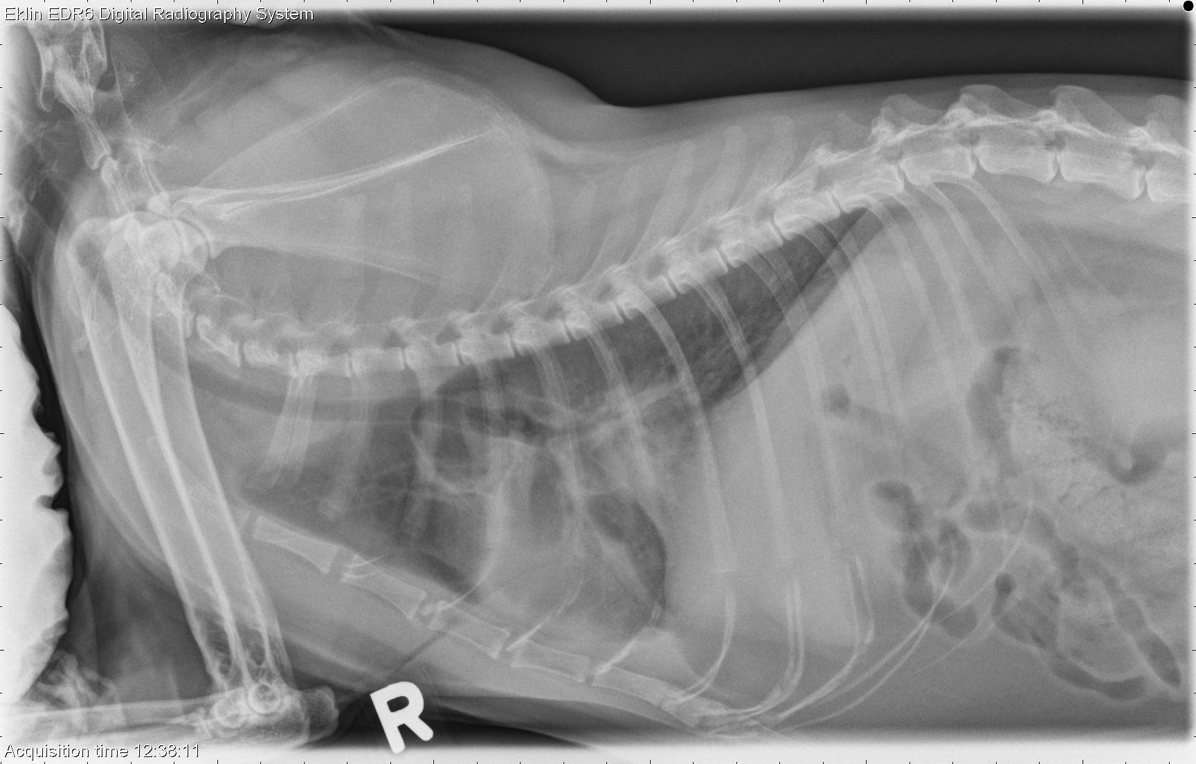
R LAT Thorax
Findings
The cardiac silhouette is normal in size and shape, and the pulmonary vasculature is normal. There is an increased soft tissue opacity in the region of the accessory lung lobe or caudal mediastinum, that silhouettes with the diaphragm and obscures the caudal cardiac silhouette. Soft tissue opacity fissure lines are visible between several lung lobes. There is a mild bronchial pattern throughout the lungs. No significant abnormalities are seen in the musculoskeletal structures.
DDx
- Mass in the accessory lung lobe or caudal mediastinum (neoplasia, granuloma, abscess)
- Pleural effusion (transudate, exudate, blood, chyle), likely secondary to the mass
- Bronchial pattern may be inflammatory or represent mild edema
Diagnosis
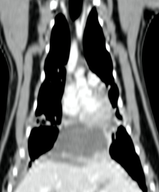
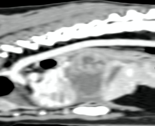
On CT images, the mass was peripherally contrast enhancing and contacted both the heart and the diaphragm. The caudal vena cava was in contact with the mass and mildly compressed, and the esophagus was dorsally displaced. Differentials remained a caudal mediastinal or accessory lung lobe mass, likely neoplastic.
The mass was found to be adhered to the pericardium and diaphragm at surgery, and was non-resectable. Necropsy diagnosis was accessory lung lobe adenocarcinoma with extension to the diaphragm and epicardium.