Intestinal Foreign Body
Publication Date: 2010-11-29
History
2 year old cross breed with three day history of lethargy, anorexia and two bouts of vomiting.
3 images
Findings
There is marked focal dilation of multiple small intestinal loops which contain geometrically arranged radiolucent material. The colon contains gas and is corrugated. The peritoneal detail is decreased and the abdomen is moderately distended. The spleen has an irregular margin with a protruding nodule on the right lateral projection. There is spondylosis deformans in the lumbar spine, and multiple metal fragments are visible in the pelvic tissues.
DDx
Small intestinal mechanical obstruction, possibly by corn cob. The poor peritoneal detail may indicate effusion due to inflammation or peritonitis. Splenic nodule may be due to lymphoid hyperplasia or neoplasia. Spondylosis deformans and metallic foreign bodies in the soft tissues are incidental.
Ultrasound
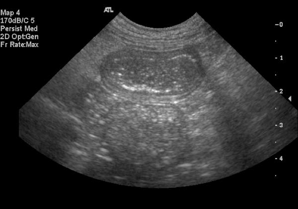
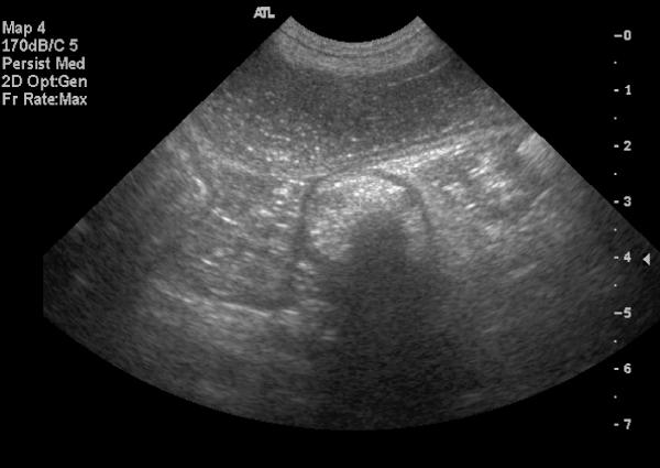
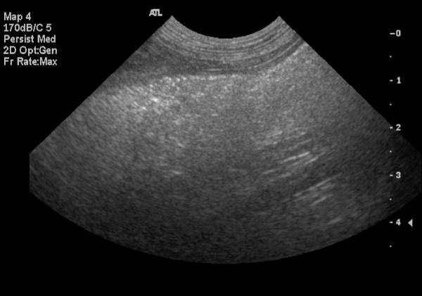
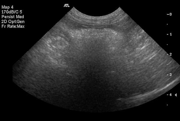
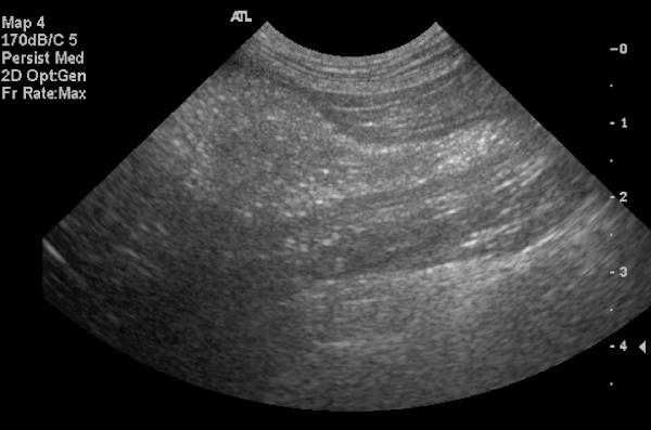
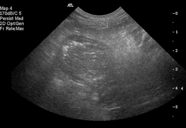
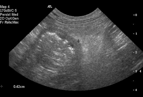
There are multiple fluid distended loops of small intestine, one of which contains foreign material with acoustic shadowing. The small intestinal wall is thickened in this region with surrounding hyperechoic mesentery and a small amount of free fluid. There is mild mesenteric lymphadenopathy. Two masses are present within the spleen.
Mechanical foreign body obstruction with local enteritis and possible peritonitis. Probable reactive mesenteric lymphadenopathy. Splenic nodules may be lymphoid hyperplasia or neoplasia.
Diagnosis
Two corn cob foreign bodies were found on exploratory laparotomy and removed via enterotomy. Septic peritonitis was present. The splenic masses were EMH and nodular hyperplasia.