Hypereosinophilic Syndrome
Publication Date: 2010-11-22
3 images
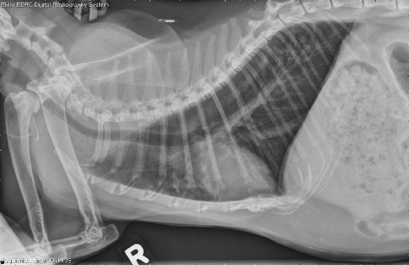
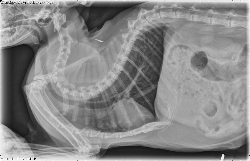
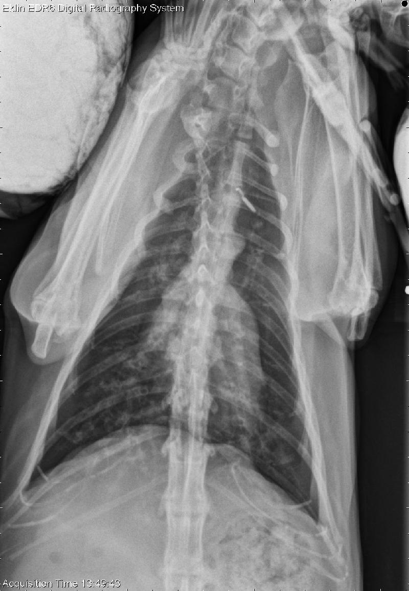
Findings
There is a multifocal mixed pulmonary pattern within the lungs with a primarily bronchial component. There is also enlargement of the pulmonary arteries with moderate cardiomegaly. Within the viewable abdomen, the liver is moderately enlarged and there is a moderate amount of gas present throughout the small intestine.
DDx
- Patchy multifocal pulmonary infiltrates; differentials include granulomatous disease such as fungal pneumonia, eosinophilic bronchopneumonopathy or heart worm disease. Pulmonary neoplasia should also be considered for this patient. Thoracic ultrasound or CT is recommended if clinically indicated.
- Cardiomegaly.
- Hepatomegaly; Enlarged pulmonary arteries may indicate pulmonary hypertension or heartworm disease.
Diagnosis
Bronchoscopy with tracheal lavage and cytology revealed moderate eosinophilic inflammation with previous/chronic hemorrhage. The presumptive diagnosis was hypereosinophilic syndrome.
Additional Images


After one month of steroid and antibiotic treatment, the pulmonary infiltrates were markedly improved.