Hiatal Hernia
Publication Date: 2010-11-22
History
7 year old female neutered English Bulldog. Found in the evening laterally recumbent and minimally responsive. No known trauma.
5 images
Findings
A complete study of the thorax with the addition of a VD view is available which was performed after-hours. A small amount of pleural effusion is present. The cardiovascular and pulmonary structures are within normal limits for the breed, age and body condition of the patient. On the right lateral and one of the VD views there is a well marginated, ovoid soft tissue structure extending into the thorax from the dorsal diaphragm on midline. The trachea is diffusely, mildly narrowed and undulating throughout its length. Within the viewable abdomen there is diffuse loss of serosal detail with a wispy opacity throughout.
Diagnosis
- Mild pleural effusion
- Sliding hiatal hernia
- Mild hypoplastic trachea compatible with the breed
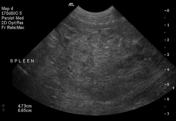
- The appearance of the abdomen is consistent with the previously diagnosed hemoabdomen
The splenic mass was surgically removed (hemangiosarcoma), and a gastropexy was performed to correct the hiatal hernia.
