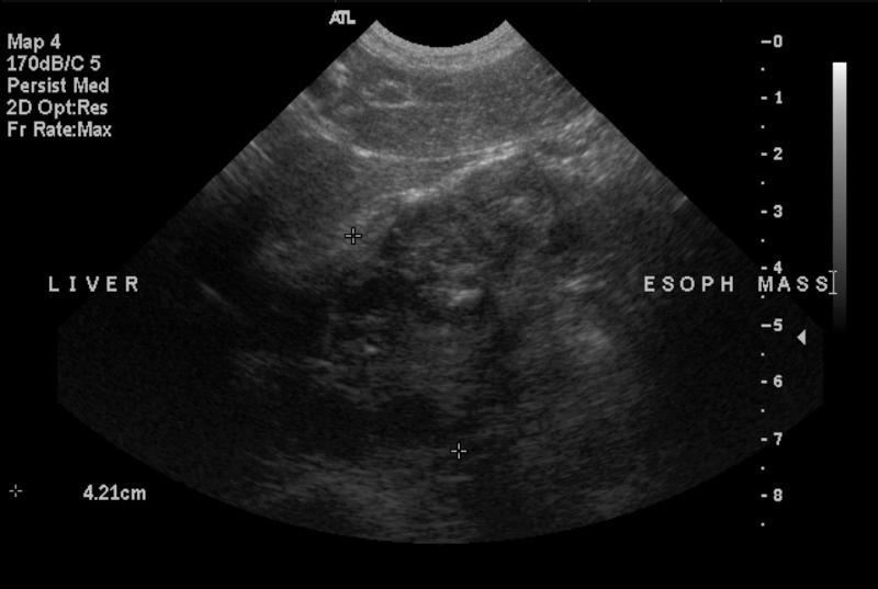
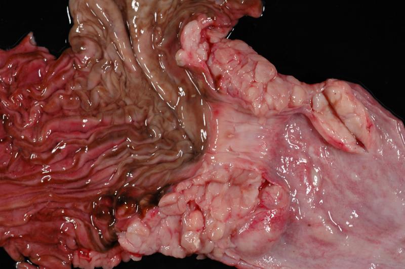
Leiomyosarcoma
Publication Date: 2010-06-13
History
13 year old MN Rottweiler. Regurgitation with increased frequency over the last 6 months, now daily.
2 images
Findings
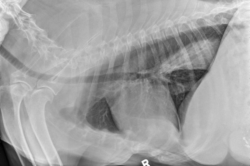
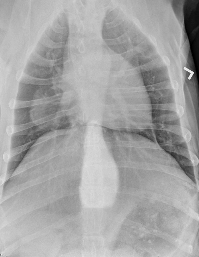
Radiographs: The caudal intrathoracic esophagus is distended and filled with soft tissue opacity. In addition, there is a fat dense mass of tissue best noted on the DV projection just lateral to the cardiac silhouette. The rest of the pulmonary parenchyma is within normal limits. The cardiovascular structures are also within normal limits. There is granular mineral debris within the gastric lumen, and the liver has rounded borders.


