Hemangiosarcoma
Publication Date: 2010-06-12
History
12 year old male neutered Golden Retriever. Owner noticed left thoracic wall mass.
3 images
Findings
Radiographs: There is soft tissue density which is well-defined on the lateral views and more poorly on the DV view, which is overlying the left cranial lung lobe. There appears to be compression of this lung lobe. The ventral aspect of the left 3rd rib is unable to be visualized. A soft tissue mass extends laterally from this point. Multiple small soft tissue densities are present primarily ventrally. The cardiovascular and the remaining pulmonary structures appear within normal limits.
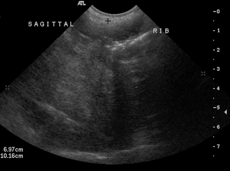
Ultrasound: Ultrasound exam of the mass lesion on the thoracic wall shows a very heterogeneous mass lesion measuring at least 6.3 cm by 7.4 cm. Within the mass lesion, there are more hypoechoic areas and hyperechoic areas with acoustic shadowing. it is visualized that the mass originates from a rib as was previously mentioned on thoracic radiographs. There is a very small amount of pleural effusion visualized and some areas of the lung surface appears somewhat irregular. There is no focal lesion visualized.
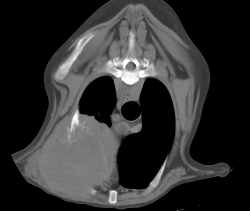
CT: Contiguous 5-mm slices of the thorax were obtained. There is an approximately 8 cm diameter rounded expansile soft tissue mass in the left cranial thorax centered at the third rib. There is near complete destruction of the rib with amorphous mineralization of the soft tissue mass. The mass invades the thoracic cavity and lies adjacent to the heart base and great vessels. There are numerous small soft tissue nodules throughout all lung lobes. There is new bone production on the medial ventral aspect of the left fourth rib. There is also a 1 cm thin-walled bulla in the dorsocranial aspect of the right caudal lung lobe.
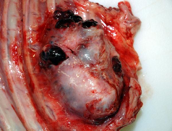
Additional Images