Nasal Foreign Body
Publication Date: 2010-04-04
History
7 year old female neutered Boxer. Nasal congestion and difficulty breathing.
4 images
Findings
THORAX A single lateral projection of the neck was obtained. There are two rounded soft tissue opacities present within the nasopharynx which appear attached to the dorsal nasopharyngeal wall. There is extension of the hyoid apparatus and abnormal air distention of the caudal nasopharynx. There is mild gasseous distention of the cervical esophagus. Within the thorax, the cardiopulmonary structures appear within normal limits.
DDx
RADIOGRAPHIC IMPRESSIONS Soft tissue masses within the nasopharynx.
CT and rhinoscopy are recommended if indicated. Unremarkable thorax without evidence of metastatic disease.
Diagnosis
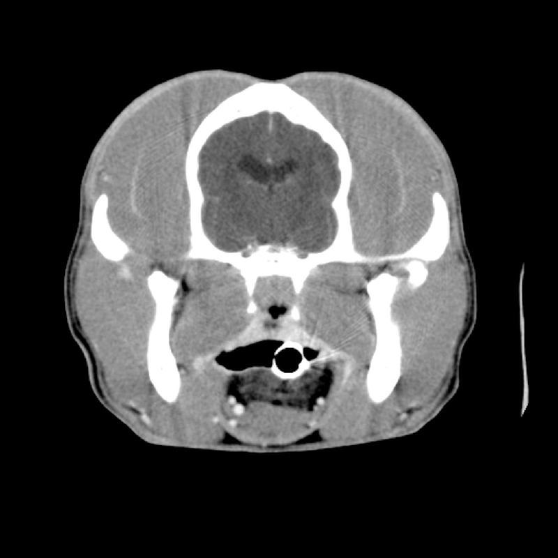
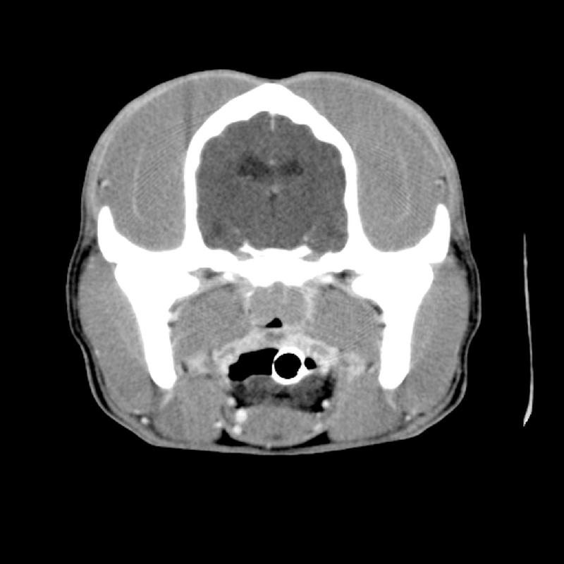
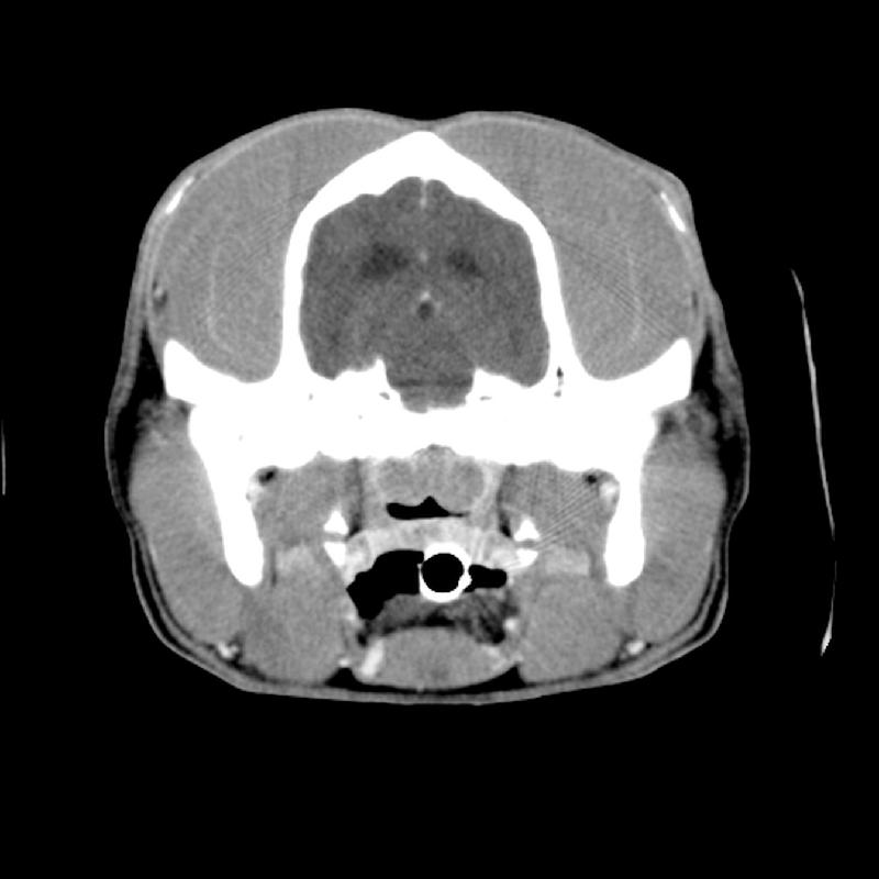
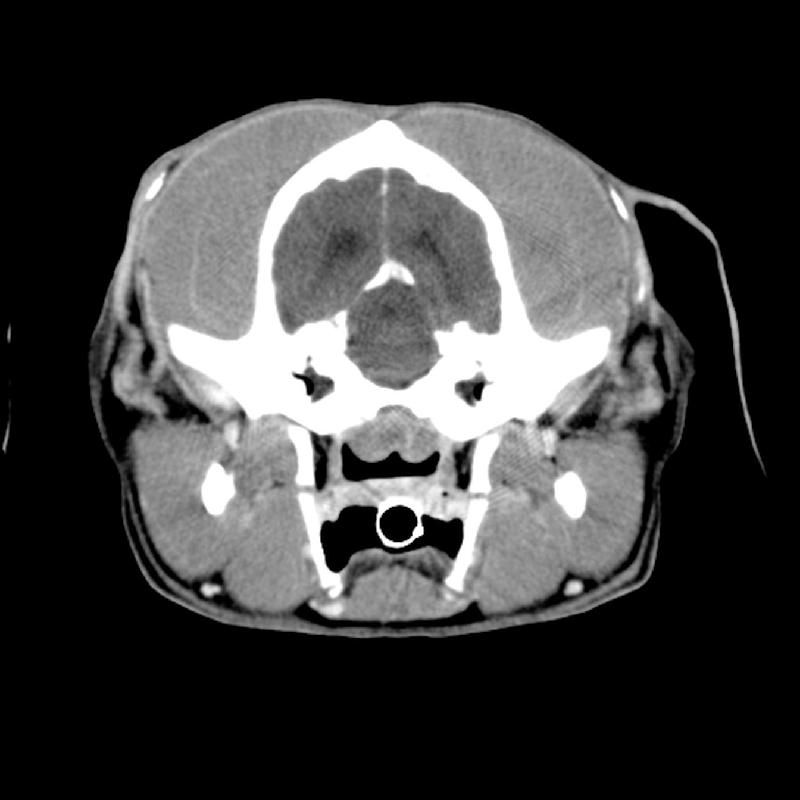
- 2 caudal nasopharyngeal masses were seen on CT
- The masses were identified and biopsied on rhinoscopy
- Marked lymphocytic, plasmacytic, and histiocytic pharyngitis with intralesional foreign body