Linear Foreign Body
Publication Date: 2010-01-22
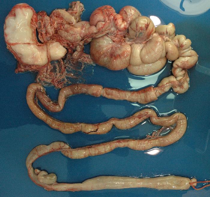
History
Young adult male intact Terrier cross that presents for evaluation of emaciation, inability to gain weight and panhypoproteinemia. The owner had adopted the dog from a shelter several weeks earlier after he had been found in an emaciated state. No weight gain to date.
3 images
Findings
There is severe loss of serosal detail due to the thin body condition of the patient. Multiple loops of small bowel are severely gas distended and appear to have a plicated pattern. The stomach is full of granular appearing ingesta which is also seen extending down the duodenum on the v/d projection. An angular mineral opacity is present in the caudal small bowel. There is a convex soft tissue density seen intruding into an enlarged loop of small bowel in the dorsal abdomen on the left lateral projection.
DDx
Distended bowel loops with a convex soft tissue appearance could indicate intussusception. The foreign material in the stomach and duodenum, gravel sign, and plication suggest a linear foreign body and chronic partial obstruction. Decreased dorsal detail is likely in part due to poor body condition although the presence of peritoneal effusion is likely.
Discussion
The plication pattern is difficult to discern because of the marked distension of the bowel loops. Multiple intussusceptions containing hard-shadowing foreign material were seen on ultrasound exam. On radiographs, the intussuscepting bowel is often outlined by gas in the distal portion of the bowel loop, appearing as a blunt-ended, soft tissue opacity.