Eosinophilic Bronchopneumopathy
Publication Date: 2010-01-13
History
10 year old female neutered Cocker Spaniel with chronic, but recently worsening cough. Previous left cranial lung lobectomy.
3 images
Findings
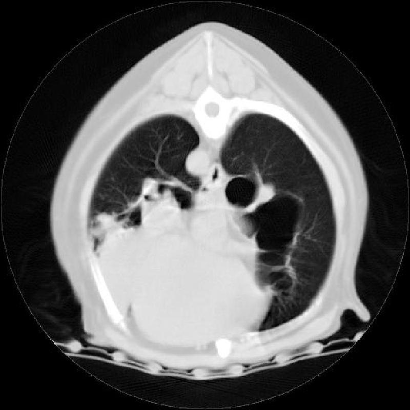
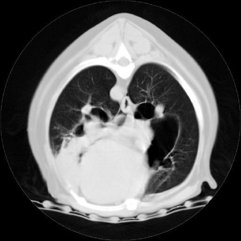
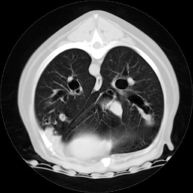
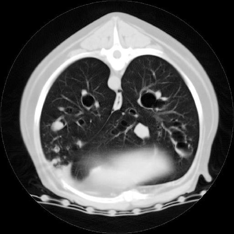
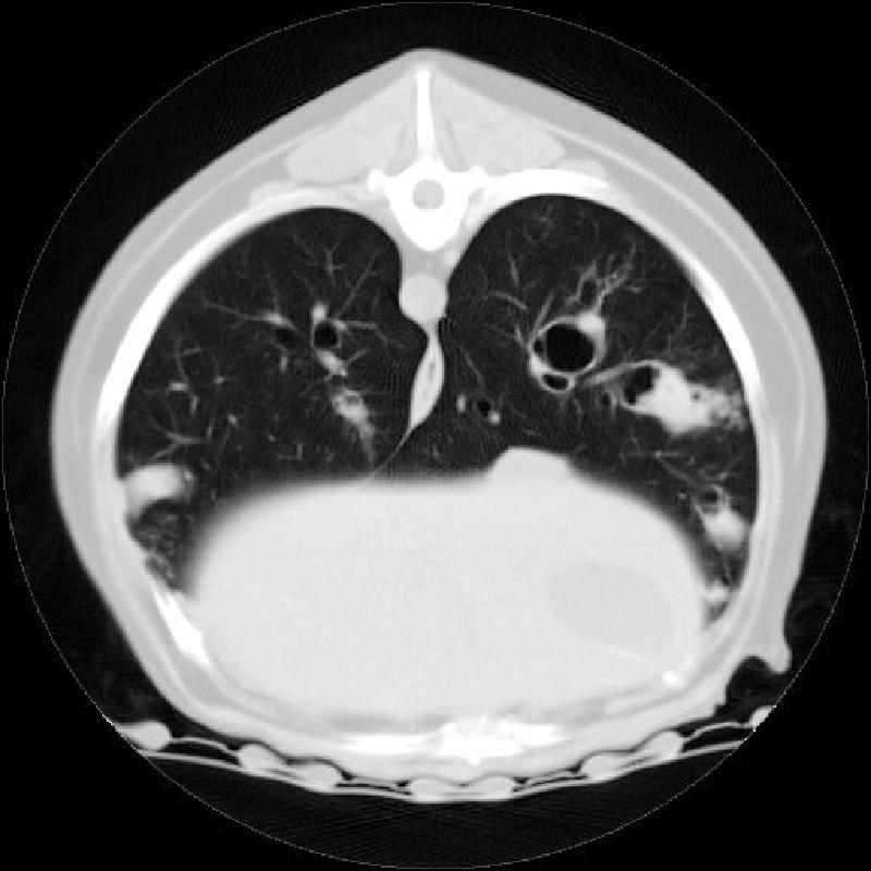
There is a severe bronchiectasis of the right middle lobar bronchus. This appears as sacculated, thin-walled mineral opacities, similar to bullae. There is an alveolar pattern in the right cranial and other lung lobes associated with mild dilation and poor tapering of the bronchi. The cardiovascular structures appear within normal limits. Two hemoclips are visible in the left thorax. There is a mild bronchial pattern throughout the lungs.
Discussion
Eosinophilic bronchopneumopathy (previously called PIE) is a cause of severe bronchial inflammatory disease. In this dog, the chronic changes and secondary inflammation and pneumonia necessitated lung lobe removal and caused recurring infiltrates. The CT images below show the dramatic dilation of bronchi, the thickened walls visible on radiographs, and the infiltrates in the lungs.
