Emphysematous Cystitis
Publication Date: 2010-01-12
Findings
The pylorus appears mildly enlarged on the lateral projection, and is filled with fluid and granular material. The small intestine is uniform in diameter.
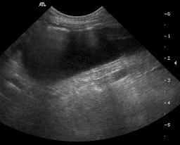
There are several irregular gas lucencies in the urinary bladder, best seen on the lateral projection. No calculi are visible. The kidneys are normal in size and shape.
Discussion
Emphysematous cystitis is commonly associated with diabetics since the glucose in the urine forms a hospitable environment for gas-producing bacteria. On ultrasound, there were multiple polyps associated with the dependent mucosa. Gas was visible in the bladder wall and free in the lumen. The urine culture resulted in growth of two types of hemolytic E. coli.


Petite A, Busoni V, Heinen MP, et al. Radiographic and ultrasonographic findings of emphysematous cystitis in four nondiabetic female dogs. Vet Radiol Ultrasound 2006;47:90-93.
Aizenberg I, Aroch I. Emphysematous cystitis due to Escherichia coli associated with prolonged chemotherapy in a non-diabetic dog. J Vet Med B Infect Dis Vet Public Health 2003;50:396-398.
Besley WM. What is your diagnosis? Emphysematous cystitis. Journal of Small Animal Practice 2004;45:283, 325.
2 images