Hemangiosarcoma
Publication Date: 2009-12-08
Patient
| Age: | 11 years |
| Sex: | male |
| Species: | Canine |
| Breed: | Golden retriever |
History
11 year old male Golden Retriever with forelimb lameness.
3 images
Findings
There is soft tissue density which is well-defined on the lateral views and more poorly on the DV view, which is overlying the left cranial lung lobe. There appears to be compression of this lung lobe. The ventral aspect of the left 3rd rib is unable to be visualized. A soft tissue mass extends laterally from this point. Multiple small soft tissue densities are present primarily ventrally. The cardiovascular and the remaining pulmonary structures appear within normal limits.
Impressions
Extrapleural mass arising from the left third rib that is invading the thoracic cavity and compressing the left cranial lung lobe. Small soft tissue nodules are suspicious for metastatic nodules.
DDx
- primary bone tumor with pulmonary metastasis (chondrosarcoma, osteosarcoma, fibrosarcoma)
- metastatic tumor to bone and pulmonary parenchyma (carcinoma, sarcoma)
Diagnosis
Additional Images
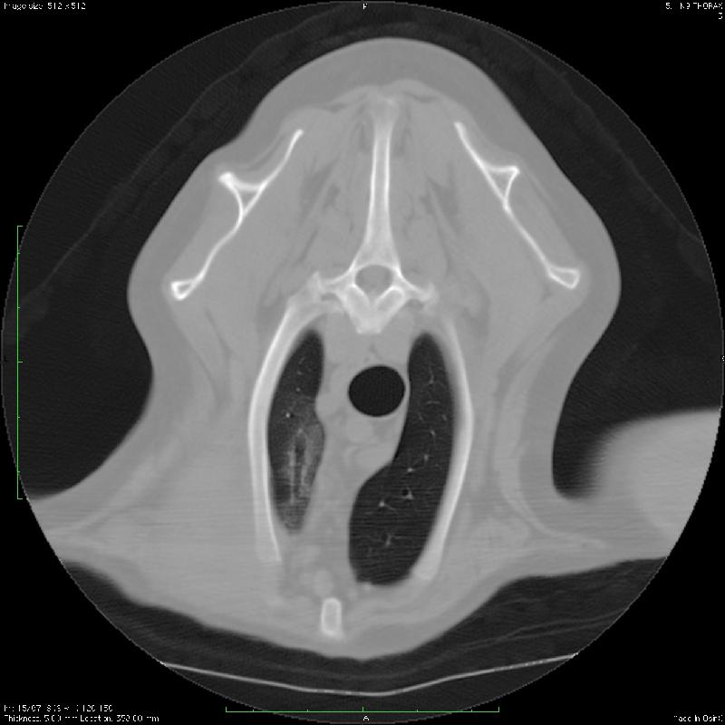
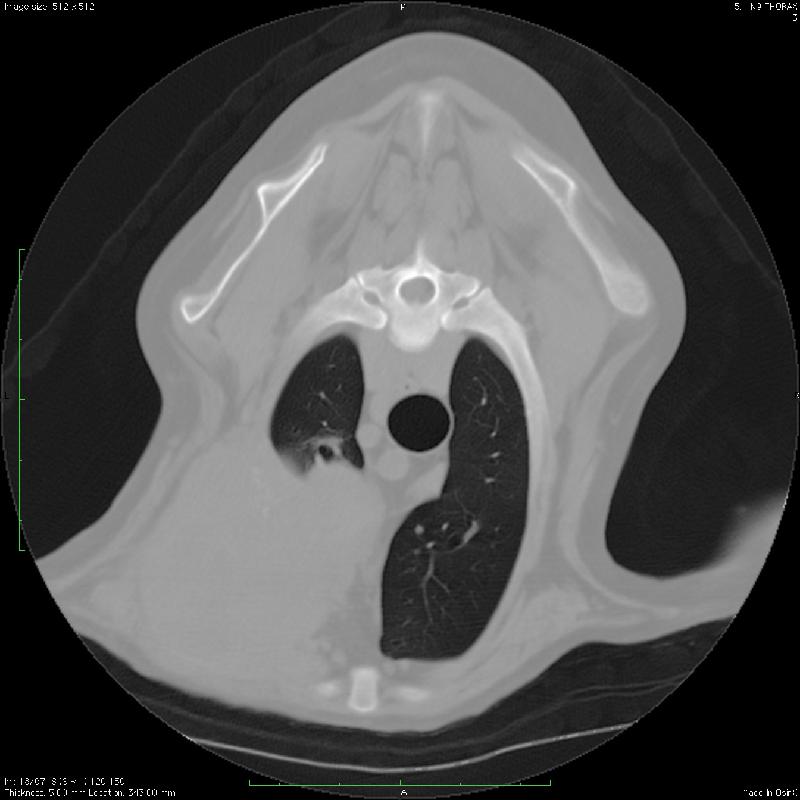
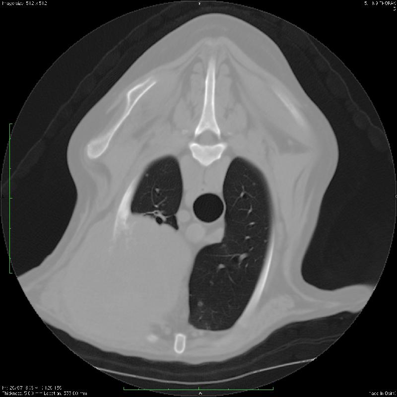
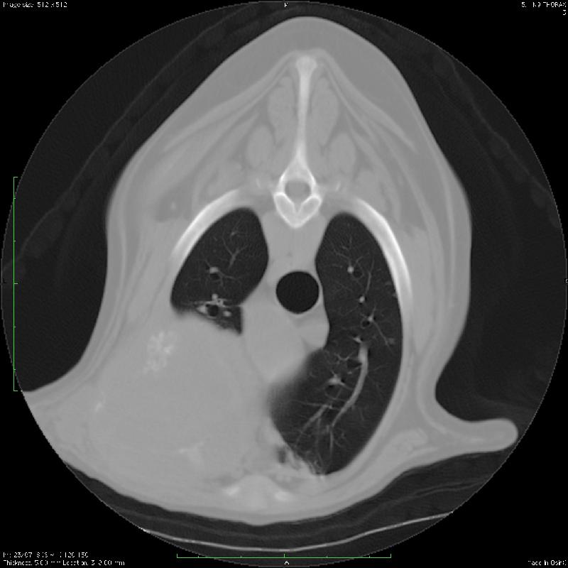
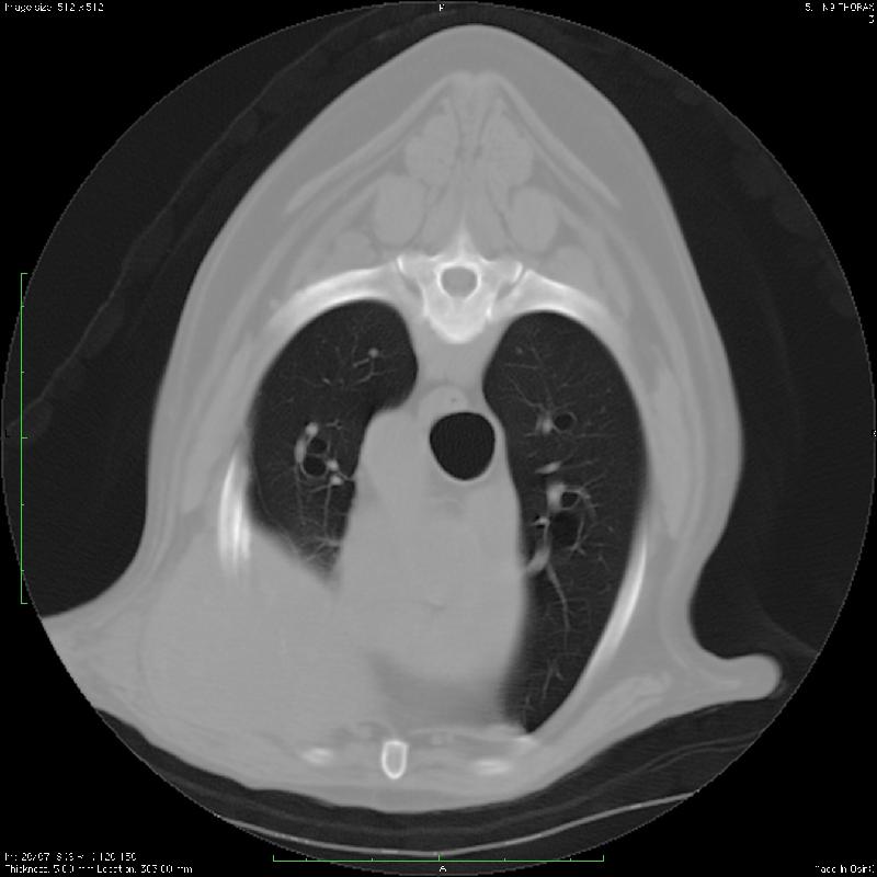
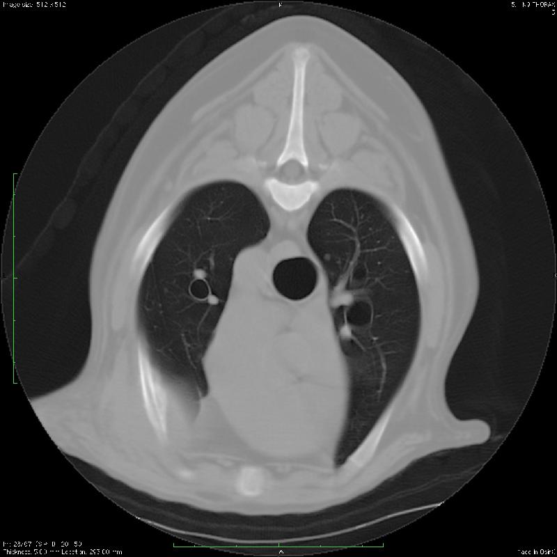
There is an approximately 8 cm diameter rounded expansile soft tissue mass in the left cranial thorax centered at the third rib. There is near complete destruction of the rib with amorphous mineralization of the soft tissue mass. The mass invades the thoracic cavity and lies adjacent to the heart base and great vessels. There are numerous small soft tissue nodules throughout all lung lobes. There is new bone production on the medial ventral aspect of the left fourth rib.
Discussion
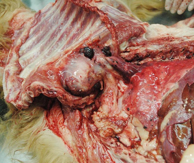
At the time of diagnosis, there were two small splenic nodules on abdominal ultrasound, and the rest of the abdomen was unremarkable. At necropsy several months later, there was extensive abdominal involvement including liver and spleen.
Files