Cecal Impaction
Publication Date: 2009-04-28
History
Long Eared Owl with historical wing amputation. Recent anorexia and lethargy, possible ataxia.
4 images
Findings
On survey radiographs, the right wing amputation is evident. The bird's body condition is thin. The heart appears mildly enlarged.
There is a large, soft tissue opacity mass occupying the majority of the celom. The proventriculus is displaced ventrally, and the ventriculus is not clearly seen.
7 mL 50% barium suspension was used to perform an upper GI exam. This revealed a ventrally displaced proventriculus, and the intestines wrapped peripherally around the mass. The brush border visible on the mucosal surface is normal. The intestinal tract appears to be patent with barium present in the cloaca at 30 min.
Diagnosis
Cecal impaction.
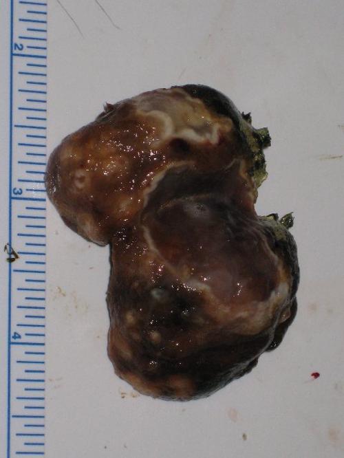
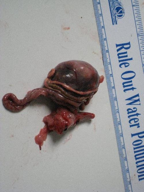
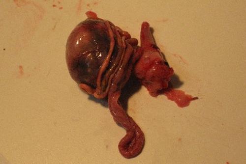
Pathology
Necropsy revealed thin bird. The gi tract appeared to be empty. There was a 2.5” diameter mid coelomic mass with intestines wrapped and adhered to the surface. The mass was located at the distal tip of the L cecum. The surface of the mass had ecchymoses that from fresh to old (brown) with several 3-5 mm white areas that appeared to be fungal colonies that could be seen through the wall of the cecum. On cut surface the mass was filled with brown firm oily fecal material that has an oily consistency. The surface of this material had several areas that appeared to be fungal placques. The corresponding surface on the cecum wall appeared ulcerated.
Microscopic interpretation: Cecum: Diffuse moderate lymphoplasmacytic and mild erosive typhlitis.
Comments The visualized cecal mass appears to be a distended and impacted cecum, evidence of neoplasia if not detected in the examined tissues. There is evidence of a chronic inflammatory process involving the mucosal surface, with mild erosion of the cecal lining epithelium. A specific etiology for this apparent cecal stasis, however, is not evident in the examined sections.