Hydronephrosis
This work is licensed under the Creative Commons Attribution-Noncommercial-No Derivative Works 3.0 United States License. To view a copy of this license, visit http://creativecommons.org/licenses/by-nc-nd/3.0/us/ or send a letter to Creative Commons, 171 Second Street, Suite 300, San Francisco, California, 94105, USA.
Publication Date: 2008-12-09
History
9 year old male neutered mixed-breed dog. Acute paraparesis with tentative diagnosis of T3-L3 myelopathy.
One lateral projection was obtained pre-myelogram, and two additional abdominal radiographs were obtained post-myelogram and hemilaminectomy.
3 images
Findings
On the initial projection of the abdomen, the stomach is moderately distended with ingesta. On all projections of the abdomen, a large soft tissue mass affect is present in the central abdomen resulting in displacement of the small intestines caudally and laterally. A large, irregular mineral opacity is present in the dorsal, right lateral abdomen at the level of L2. On the lateral projections of the abdomen, a gastric tube is present. Skin staples are present along dorsal mid-line from T10 to L4-5. The articular facets at T12-13 and T13-L1 are absent.
The left kidney and the spleen are visible and appear normal. There is a small amount of contrast in the urinary bladder and in the subarachnoid space secondary to myelography.
Additional Images
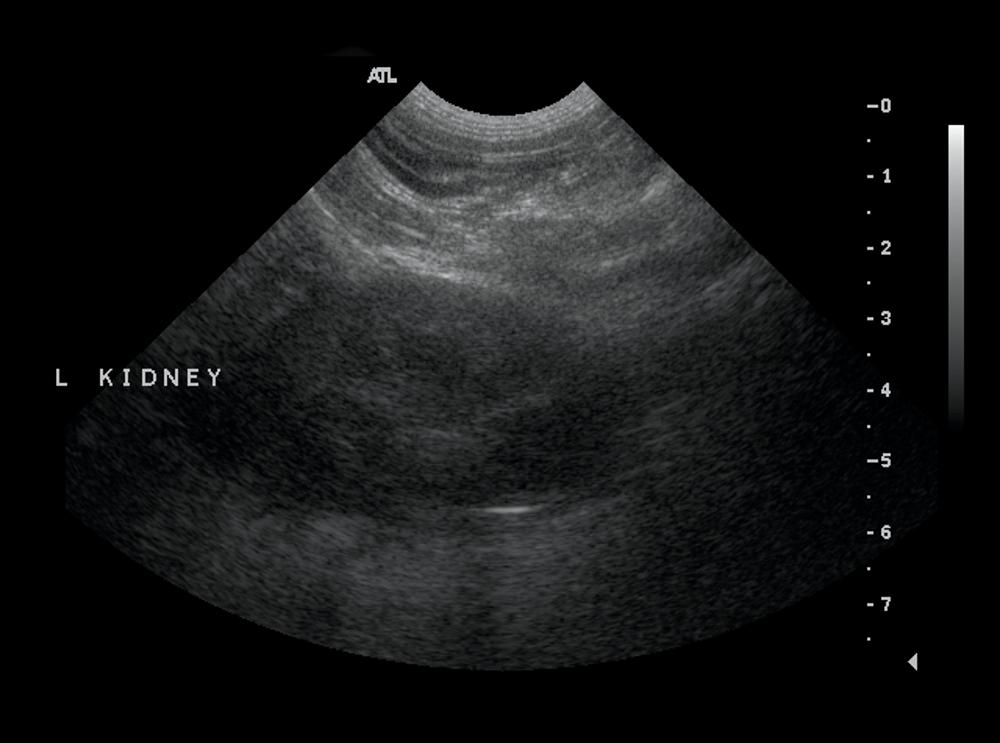
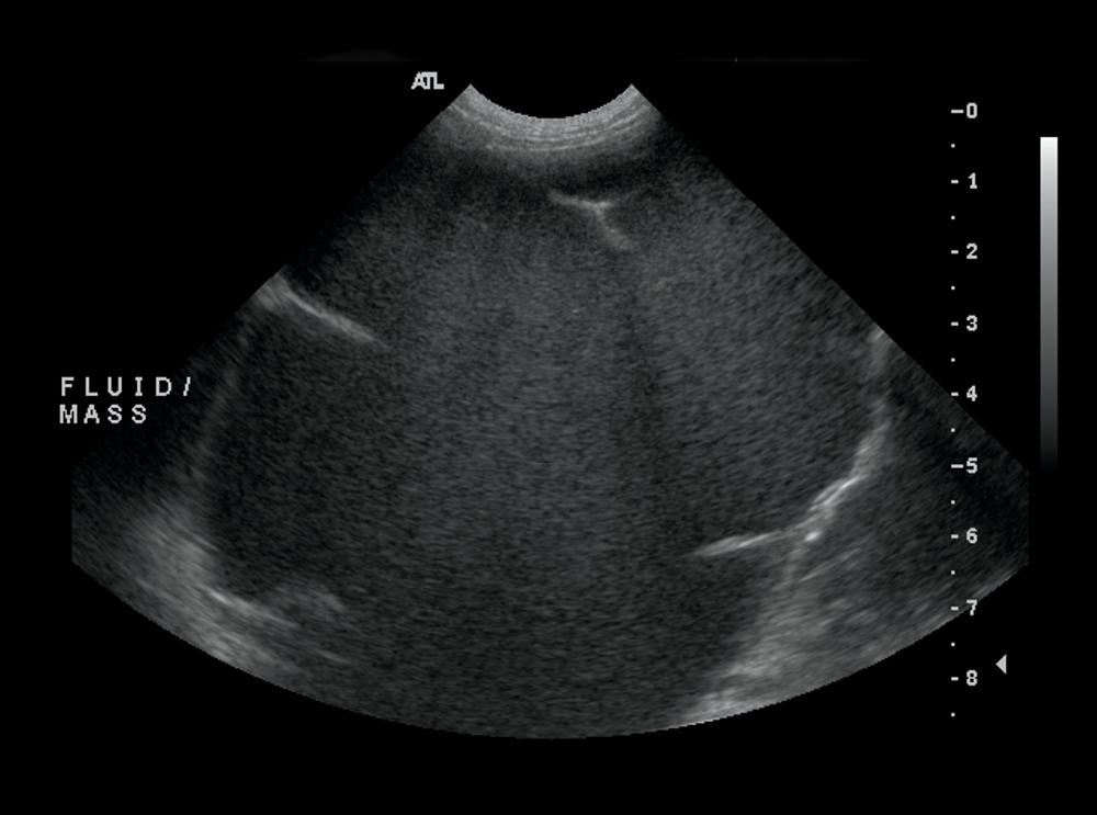
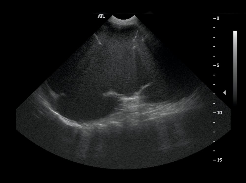
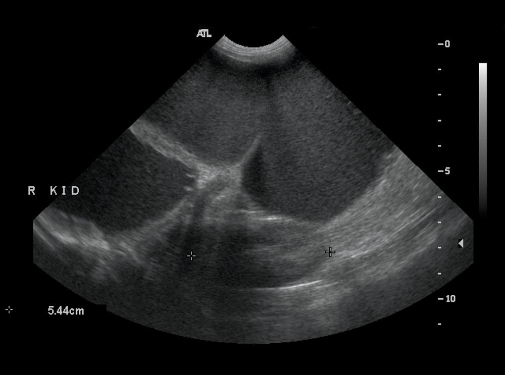
On ultrasound, the large, fluid-filled mass had multiple septae which are characteristic of a hydronephrotic kidney, and represent the connective tissue and arcuate arteries. The kidney was located in the central abdomen, so it was difficult to determine whether it was right or left. The left kidney is visible in the far field on the final image, so the hydronephrosis involved the right kidney.


Diagnosis
Right renal hydronephrosis, most likely secondary to ureteral obstruction.
Discussion
This very large abdominal mass is causing such a great mass effect that it is difficult to determine its origin. The typical signs of right renal enlargement such as ventral displacement of the ascending colon and cecum are not clearly evident. However, no normal right kidney is visible. The mineralization in the dorsal abdomen is also most likely associated with the right renal pelvis.
Because the immediate problem for this patient was a herniated disc, the kidney was not addressed at this time. This condition should be monitored for progression or acute disease.