Pneumothorax
This work is licensed under the Creative Commons Attribution-Noncommercial-No Derivative Works 3.0 United States License. To view a copy of this license, visit http://creativecommons.org/licenses/by-nc-nd/3.0/us/ or send a letter to Creative Commons, 171 Second Street, Suite 300, San Francisco, California, 94105, USA.
Publication Date: 2008-10-10
History
8 year old male neutered Siberian Husky who was hit by a car several hours ago.
3 images
Findings
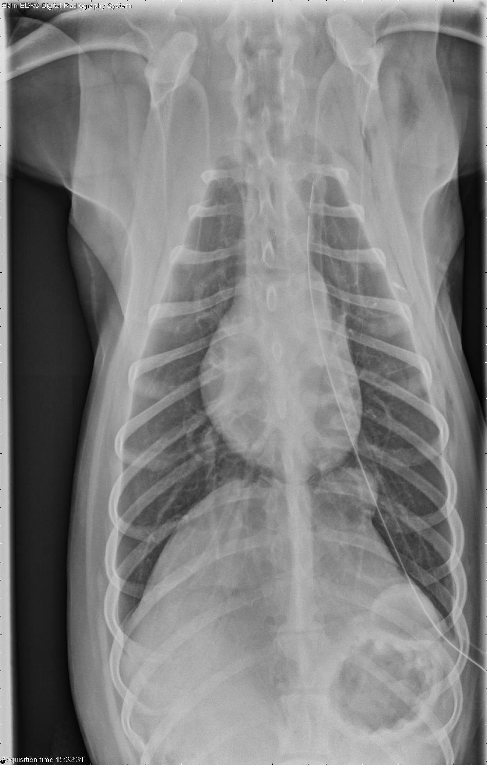
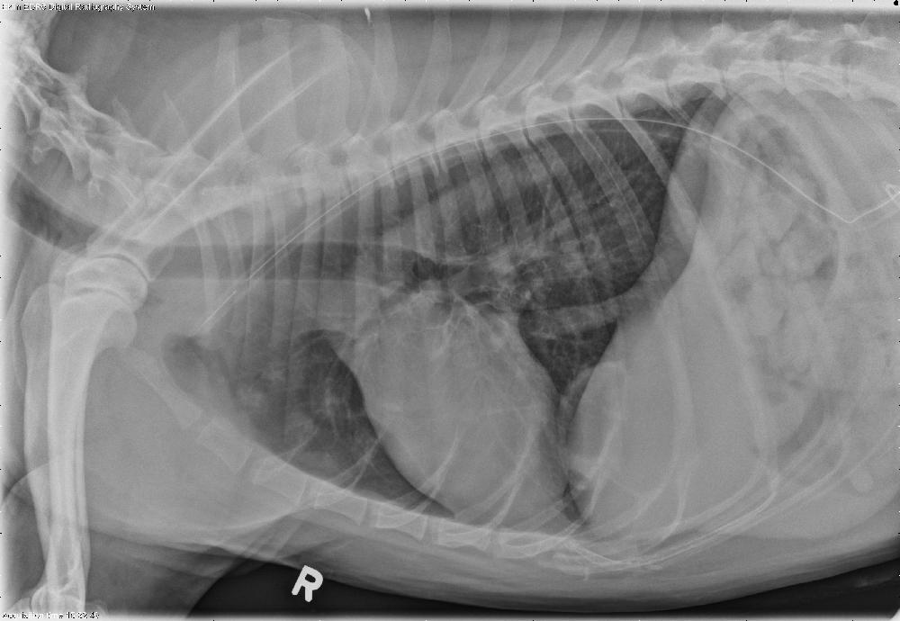
A chest tube is present terminating at the level of the first rib in the left cranial lung fields. Pneumothorax is present, worse on the right than the left. Pneumomediastinum is also evident particularly caudally. Two bullae are identified in the left caudal lung lobe, best seen on the right lateral projection and the v/d projection (click on annotations). There is an interstitial to alveolar pattern in the left cranial and left caudal lung fields.
Diagnosis
- Traumatic pulmonary bullae
- Bilateral pneumothorax
- Pneumomediastinum
- Probable pulmonary contusions (left)
Additional Images
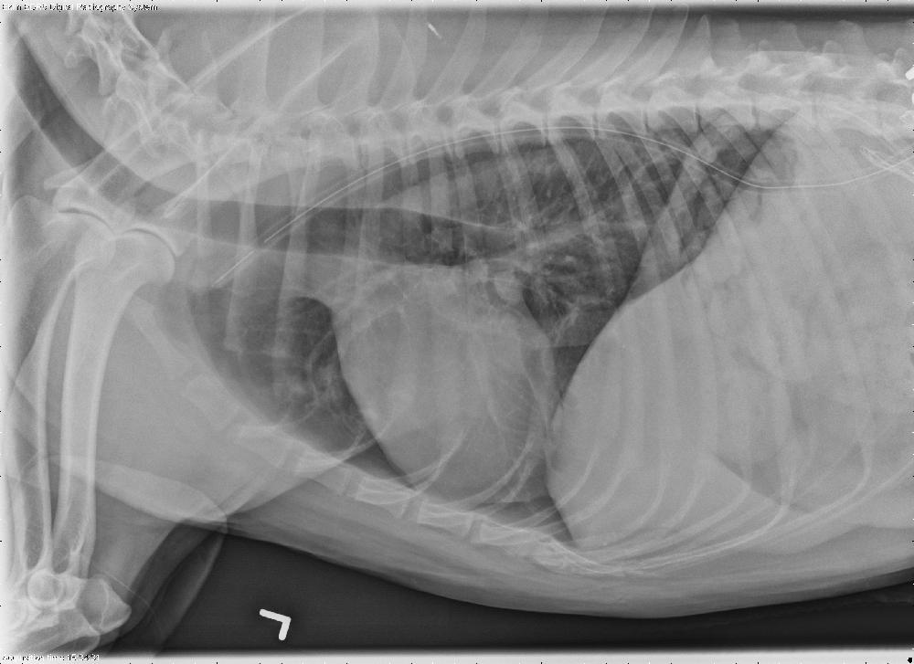
Additional radiographs were taken two days after presentation. The pneumothorax has resolved, and the chest tube remains in place. The more dorsal bulla has filled with fluid and appears as a solid soft tissue opacity. The more ventral bulla has a thick, irregular wall. These changes indicate filling of the bullae with blood or inflammatory fluid.
The dog was discharged the next day after removal of the chest tube and was doing well.