Hemangiosarcoma
This work is licensed under the Creative Commons Attribution-Noncommercial-No Derivative Works 3.0 United States License. To view a copy of this license, visit http://creativecommons.org/licenses/by-nc-nd/3.0/us/ or send a letter to Creative Commons, 171 Second Street, Suite 300, San Francisco, California, 94105, USA.
Publication Date: 2007-04-16
History
10 year old male neutered Cocker Spaniel with progressive anemia, now 16%.
Findings
There are two masses in the abdomen. The first is visible on the lateral and v/d, between the left kidney and the fundus of the stomach. The second is in the caudoventral portion of the abdoman, and is only visible on the lateral projection.
The dorsal mass is in the location of the proximal extremity of the spleen, and the distal mass is in the location of the distal extremity. The more dorsal mass is causing a mass effect and displacing the left kidney caudally (compare to right kidney)
Additional Images
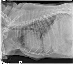
There are diffuse soft tissue opacity nodules throughout the lungs, indicating metastatic disease.
Diagnosis
Multiple splenic tumors - hemangiosarcoma with metastatic disease to the lungs.
Discussion
The location of the masses are key to identifying splenic origin. The presence of nodules on the edge of the abdominal films should prompt you to take thoracic radiographs. Metastatic disease places neoplasia at the top of the list of differential diagnoses. The anemia could have been caused by hemorrhage, RBC destruction in the spleen, or anemia of chronic disease. There was no evidence of effusion on these radiographs.
2 images