Transitional Cell Carcinoma
This work is licensed under the Creative Commons Attribution-Noncommercial-No Derivative Works 3.0 United States License. To view a copy of this license, visit http://creativecommons.org/licenses/by-nc-nd/3.0/us/ or send a letter to Creative Commons, 171 Second Street, Suite 300, San Francisco, California, 94105, USA.
Publication Date: 2007-03-19
History
11 yr female neutered Golden Retriever with two month history of straining to urinate. Lameness of right pelvic limb for 1 week.
Findings
On radiographs of the pelvis, there is a soft tissue opacity mass dorsal to the colon in the retroperitoneal space. The mass is in the region of the sublumbar lymph nodes, and is deviating the colon ventrally.
The right ilium is irregular on both views, with an expansile area visible on the lateral projection. Several oblique positions of the v/d were required to highlight the affected area.
The dorsal wall of the bladder has an unusual concave shape, giving the appearance of a mass effect between the colon and the bladder. However, on ultrasonographic examination there was nothing abnormal in this area.
Diagnosis
Urethral transitional cell carcinoma with metastasis to local lymph nodes and pelvis.
Discussion
Urethral transitional cell carcinoma occurs more often in female dogs. The urethra becomes progressively thickened, which can be seen on ultrasound exam if the lesion is cranial to the pelvis. The urethra often appears hypoechoic with a hyperechoic line at the mucosal surface. The tumor may extend to the trigone of the bladder, where it can cause obstructive hydronephrosis.
Urethral disease can also be diagnosed by retrograde urethrogram. It appears as irregularity or nodular wall, with possibility of contrast leakage into the periurethral tissues.
2 images
Additional Images


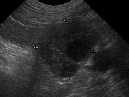
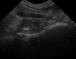
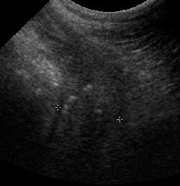
On abdominal ultrasound, the medial iliac lymph nodes were markedly enlarged and hypoechoic with cystic areas. The right ilium had an irregular surface with hypoechoic tissue next to it that contained mineralized foci. The third image shows the thickened urethra between calipers. The urethra also contains mineralized areas.
References
- Hanson JA, Tidwell AS. Ultrasonographic appearance of urethral transitional cell carcinoma in ten dogs. Vet Radiol Ultrasound 1996;36:293-299.
- Moroff SD, Brown BA, Matthiesen DT, et al. Infiltrative urethral disease in female dogs: 41 cases (1980-1987). J Am Vet Med Assoc 1991;199:247-251.
- Ticer JW, Spencer CP, Ackerman N. Transitional cell carcinoma of the urethra in four female dogs: its urethrographic appearance. Veterinary Radiology 1980;21:12-17.
- Mellanby RJ, Chantrey JC, Baines EA, et al. Urethral haemangiosarcoma in a boxer. J Small Anim Pract 2004;45:154-156.