Perinephric Pseudocysts
This work is licensed under the Creative Commons Attribution-Noncommercial-No Derivative Works 3.0 United States License. To view a copy of this license, visit http://creativecommons.org/licenses/by-nc-nd/3.0/us/ or send a letter to Creative Commons, 171 Second Street, Suite 300, San Francisco, California, 94105, USA.
Publication Date: 2007-03-12
History
13 year old female neutered domestic short haired cat with history of hematuria and hyperthyroidism.
Findings
The right kidney is markedly enlarged, with a rounded shape. It is distorting the abdominal wall on the v/d projection and displacing the ascending and transverse colon medially and ventrally. The left kidney also has an abnormal shape, with reduced length and increased width.
There is also spondylosis deformans at the L5-6 intervertebral disc space, which is an incidental finding.
Discussion
Perinephric pseudocysts are associated with chronic renal disease, but are often asymptomatic. The association between the pseudocysts and this cat's hematuria is unclear. The hematuria may have been due to pressure from the pseudocysts, or idiopathic in nature.
2 images
Additional Images
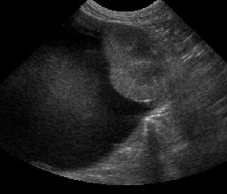
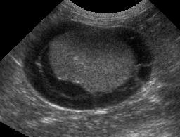
On ultrasound images, both kidneys were surrounded by large cystic structures filled with echogenic fluid. There are fine septae visible in the pseudocyst surrounding the left kidney. Drainage of both collections of fluid were clear and colorless except for the last 15 ml from the right kidney, which was hemorrhagic. Both kidneys were small and irregular with poor corticomedullary distinction.
References
- Ochoa VB, DiBartola SP, Chew DJ, et al. Perinephric pseudocysts in the cat: a retrospective study and review of the literature. Journal of Veterinary Internal Medicine 1999;13:47-55.