Intussusception
This work is licensed under the Creative Commons Attribution-Noncommercial-No Derivative Works 3.0 United States License. To view a copy of this license, visit http://creativecommons.org/licenses/by-nc-nd/3.0/us/ or send a letter to Creative Commons, 171 Second Street, Suite 300, San Francisco, California, 94105, USA.
Publication Date: 2007-03-04
History
6 month old male neutered Cairn Terrier with 3 days of vomiting and diarrhea, and progressive lethargy.
Findings
There are markedly dilated bowel loops in the caudal portion of the abdomen. More normal sized loops are gas-filled in the cranial abdomen. The peritoneal detail is poor, likely because of his young age.
DDx
Mechanical obstruction - foreign body, intussusception.
Diagnosis
Jejunal-jejunal intussusception.
Discussion
Intussusceptions are common in young dogs secondary to intestinal hypermotility. The bowel "telescopes" on itself causing a mechanical obstruction, or partial obstruction. Most intussusceptions involve the ileum and colon. It is more rare to see a jejunal intussusception. The radiographic findings fit with the diagnosis. A more proximal obstruction results in focal bowel dilation such as in this case. Distal obstruction, such as in a usual intussusception, often causes dilation of the entire proximal or oral small intestine.
3 images
Additional Images


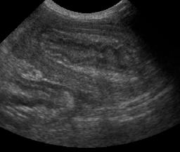
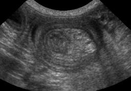
Ultrasonography diagnosed the intussusception. You can see the "bowel within bowel", or concentric rings of intestine in both the longitudinal and the cross-sectional image. There is often mesenteric fat pulled into the intussusception, which appears hyperechoic on the transverse image.