Osteosarcoma
This work is licensed under the Creative Commons Attribution-Noncommercial-No Derivative Works 3.0 United States License. To view a copy of this license, visit http://creativecommons.org/licenses/by-nc-nd/3.0/us/ or send a letter to Creative Commons, 171 Second Street, Suite 300, San Francisco, California, 94105, USA.
Publication Date: 2007-02-12
History
9 year old male neutered Rottweiler with lameness for 1 month.
Findings
There is an aggressive bone lesion in the proximal right humerus. There is lysis in the medullary cavity, with a long zone of transition. There is periosteal reaction along the cortices which extends into the soft tissues.
There is also an opacity in the right hemithorax which is adjacent to the right body wall. It is seen best on the d/v and left lateral projections. A second opacity is visible overlying the caudal lungs on this projection, and is not visible on the v/d.
DDx
Humerus - osteosarcoma, fungal osteomyelitis, metastatic disease
Diagnosis
Osteosarcoma with metastatis to rib
Discussion
The thoracic nodule was difficult to localize on the standard projections. Visibility of an extrapleural sign depends on catching it perpendicular to the x-ray beam. Centering over the lesion and collimating can help, as can rotating the dog to either side.
The humeral lesion is distal to the metaphysis, where most osteosarcomas originate. This raises the possibility of it being a metastatic lesion from another site, or a fungal lesion. The biopsy proved osteosarcoma in this case.
4 images
Additional Images
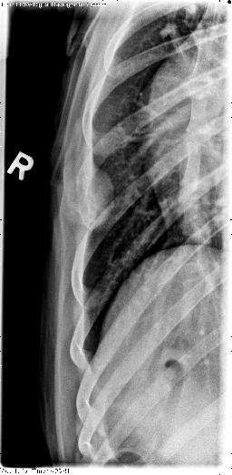
This radiograph was taken centered over the pulmonary lesion. It shows the rib has lytic and expansile components, and that the rib mass is protruding into the thorax. This is most likely metastatic disease from the osteosarcoma. It is a very nice example of an extrapleural sign, where the mass has a broad base and extends to the pleural surface.